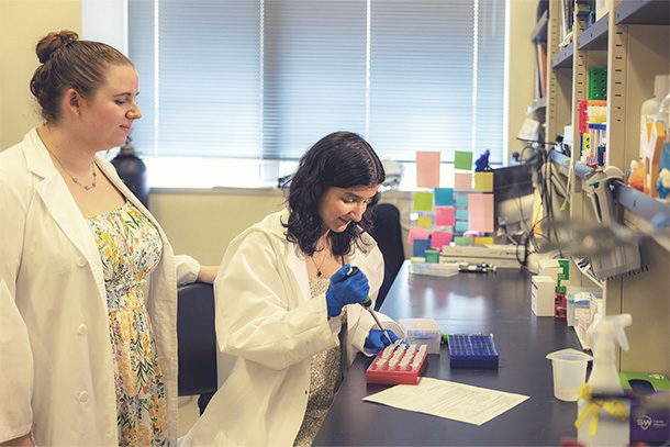
Brianna Hnath, left, is doctoral candidate in biomedical engineering at Penn State and co-author of the study. Credit: Courtesy of the Dokholyan lab / Penn State.
Toxic protein may contribute to ALS development
New study elucidates the physiological processes that may contribute to ALS development and identifies a potential therapeutic target
Oct 11, 2024
Editor's note: This article originally appeared on Penn State News.
By Christine Yu
HERSHEY, Pa. — A toxic version of a certain protein may affect brain, spinal cord and skeletal muscle tissues differently, leading to the complex development and progression of amyotrophic lateral sclerosis (ALS), according to a new study by a team of researchers from Penn State College of Medicine. The study represents a step forward in understanding the physiological processes that may give rise to ALS and identifies a potential therapeutic target for future treatments for ALS. The team published their findings in the journal Structure.
“In ALS, like other neurodegenerative diseases, there are proteins that tend to aggregate in harmful clusters. One that’s associated with ALS is superoxide dismutase 1, or SOD1,” specifically in its trimeric form,” said senior author Nikolay Dokholyan, G. Thomas Passananti Professor at the Penn State College of Medicine and professor of biochemistry and molecular biology.
Dokholyan explained that SOD1 typically exists as a dimer, a protein composed of two identical units. Under certain conditions, SOD1 will change its shape and reassemble itself into a three-unit form called a trimer.
“We need to understand how the SOD1 trimers kill cells and the mechanisms involved,” he said.
ALS is a progressive neurodegenerative disease that affects nerve cells, called neurons, in the central nervous system and leads to muscle weakness and atrophy. Mutations of SOD1 have been implicated in approximately 20% of ALS cases with a known genetic cause and a small percentage of cases with no known genetic link. Previous research has shown that SOD1 trimers appear to gain a toxic function compared to dimers. SOD1 trimers are linked with increased cell death in models of ALS but the exact molecular mechanism behind it isn’t known, Dokholyan said.
To investigate the role SOD1 trimers play in cell dysfunction and degeneration, the team examined which proteins bind with SOD1 trimers. Dokholyan explained that they introduced SOD1 trimers into three different types of mouse tissue — brain, spinal cord and muscle tissue — and observed which proteins attached to the trimers. They then compared the protein binding partners of SOD1 trimers in the three tissues with the binding partners of SOD1 dimers.
“We were trying to see if there were any new proteins that would show up interacting with this toxic protein that hadn’t been seen before,” said Brianna Hnath, doctoral candidate in biomedical engineering at Penn State and co-author of the study. “The goal was to find the potential pathways for how this SOD1 trimer could have a toxic pathway.”
The researchers found that SOD1 trimers interact with different proteins depending on the type of tissue, which they said could explain — in part — the complex and multifaceted nature of ALS.
In brain and spinal cord tissue, SOD1 trimers bind with proteins that are involved in maintaining neuron structure, function and communication between nerve cells. The team also found that SOD1 trimers activate pathways connected to cellular aging, which may contribute to neuronal dysfunction and degeneration. In muscle tissue, SOD1 trimers were found to bind with proteins involved with metabolic processes. As a result, this interaction may directly interfere with metabolism and energy production within the muscle cells.
“The fact that we were finding different hits in the three different types of tissues, instead of one uniform hit, means that there could be different mechanisms leading to cell dysfunction and death, depending on the cell type,” Hnath said.
This finding challenges the traditional notion that muscle wasting in ALS is a secondary result of motor neuron degeneration — when these neurons don’t function normally, muscle cells aren’t stimulated, which can lead to muscle atrophy, Dokholyan explained. However, the study suggests that there may also be processes within muscle cells that are disrupted by SOD1 trimers that may cause muscle cell dysfunction and death, contributing to muscle wasting and neuron death.
“Both neurons and muscle cells are affected,” Dokholyan said. “On the neuron side, it’s potentially affecting the ability of neurons to connect to muscle, while on the muscle side, it affects metabolism.”
In particular, the study identified the protein septin-7 as a binding partner for SOD1 trimers but not native SOD1 dimers. Septin-7 plays a role in essential nerve cell processes like maintaining cellular structure and communication and has been linked to ALS in previous studies. Binding with SOD1 may disrupt these functions, leading to neuron degeneration. It raises the question if addressing this interaction could slow or disrupt ALS progression, making septin-7 a potential therapeutic target, Dokholyan said.
He noted that more research is needed to further understand the potential role of SOD1 trimers in the development of ALS, how it may lead to cellular dysfunction and death, and the specific role of septin-7, which could guide the future development of potential therapies.
Other Penn State College of Medicine authors on the paper include first author Esther Choi and Congzhou Mike Sha, joint degree students in the MD/PhD Medical Scientist Training Program.
Funding from the National Institutes of Health, the National Center for Advancing Translational Sciences and the Passan Foundation supported this work.

