
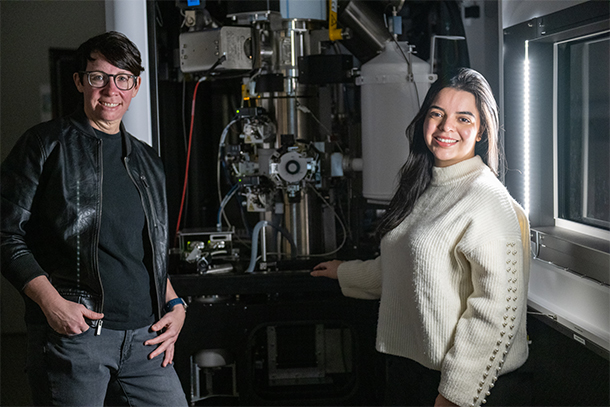
Deb Kelly (left), professor of biomedical engineering, director of the Center for Structural Oncology and Huck Chair in Molecular Biophysics; and Maria Solares, doctoral student in molecular, cellular and integrative biosciences in the Huck Institutes of the Life Sciences, stand next to the cryo-electron microscopy machine. They used cryo-EM to image individual p53 proteins isolated from brain tumor cells. Credit: Jeff Xu/Penn State. All Rights Reserved.
Guarding the genome: Researchers uncover full 3D structure of p53 protein
Understanding how tumor suppressors fail could lead to new cancer treatments
March 1, 2023
Editor’s note: One of the articles on which this story is based has been retracted. Following established research integrity processes conducted by Penn State, this paper was retracted by the journal.
By Mariah Lucas
UNIVERSITY PARK, Pa. — The tumor suppressor protein p53, known as the guardian of the genome, protects the body’s DNA from daily stress or long-term damage by triggering the cells to make repairs or to self-destruct. But mutations in the p53 gene that codes for the protein can prevent it from performing its job, making errors accumulate in the genetic code and leading to diseases like cancer.
For the first time, a Penn State-led team of researchers uncovered the complete structure of the p53 protein using patient samples. They also investigated how mutation-induced changes in the p53 structure can impact different cancers. They published their results in ChemBioChem and the International Journal of Molecular Sciences.
“We defined the full-length structure of p53, opening the door to understanding the 3D arrangements that can used to inform new therapeutics,” said Maria Solares, Penn State doctoral student in molecular, cellular and integrative biosciences in the Huck Institutes of the Life Sciences and first author on both papers. “Scientists have previously identified p53 as an important focal point in the origin of tumors, and much has been investigated on its function in cells. However, without understanding the complete structure of the p53, our knowledge of how to deal with it in diseased cells was incomplete.”
Researchers used cryo-electron microscopy (cryo-EM) to image individual p53 proteins isolated from brain tumor cells. Unique to their work, Solares said, the team used semi-conductor materials to cleanly capture the isolated p53 proteins prior to imaging. These silicon-based microchips hold the proteins in such a way that the researchers could resolve previously unseen molecular features.
“Seeing the complete structure of the p53 was like finally seeing the whole human body after only viewing limited parts or limbs for so long,” said corresponding author Deb Kelly, Penn State professor of biomedical engineering, director of the Penn State Center for Structural Oncology and Huck Chair in Molecular Biophysics. “It’s hard to understand how things work without knowledge of their entire physical makeup.”
The p53 protein comprises individual units called monomers, which combine to create larger entities, or dimers and tetramers.
“We found a mixture of monomers, dimers and tetramers were present inside cancer cells, each of which serve different purposes based on events happening inside the cell’s nucleus,” Solares said. “Results of the dimer structure revealed, for the first time, a ‘closed’ configuration of the molecule. This form of p53 is like being at the starting blocks before a race, primed and ready to run toward DNA when notified by other cellular signals that there’s a problem.”
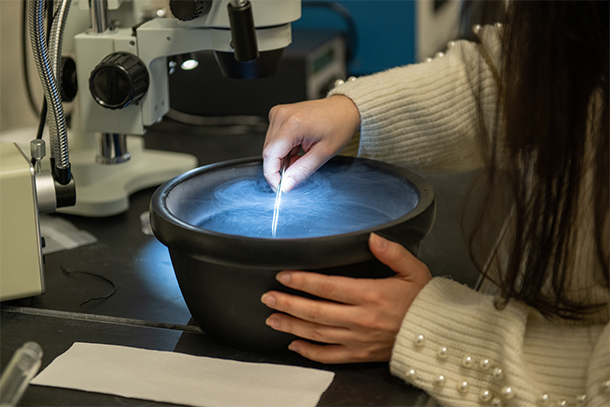
Maria Solares, first author on both papers, demonstrates the transfer of a specimen in liquid nitrogen in preparation for imaging in the cryo-electron microscopy instrument. Credit: Jeff Xu/Penn State. All Rights Reserved.
Other forms of p53, such as monomers, are considered ‘open’ because they need to bond with another unit before they can move to the starting line to perform their tasks. According to Kelly, proper communications are important to control the extent and timing of p53 deployment to the nucleus of cells, where DNA resides, and interruptions in the process are linked to the origins of diseases, including cancer.
Mutations in the p53 gene can lead to p53 proteins that are unable to communicate well, Kelly said. Once they revealed the 3D structure, the researchers examined how structural changes in p53 result in molecules that operate improperly inside cells.
“It’s well known that half of all cancers contain p53 gene mutations,” Solares said. “To take a closer look at how these mutations impact the p53 protein structure, we used molecular modeling software to simulate changes in the p53 monomer structure.”
The researchers looked at seven “hotspots,” where mutations in the protein structure are most commonly linked to cancer. These seven mutations in the p53 protein typically lead to less favorable patient outcomes in terms of disease progression and chemoresistance, Kelly said.
They found that slight shifts in the 3D structure of a mutated p53 can affect the protein’s surface charges, which work to repel and attract charges of other molecular units. According to Kelly, this can impede proper interactions between the protein and DNA, leading to a breakdown in p53’s ability to assist with regulatory or repair processes essential for maintaining healthy cells.
“Under healthy conditions, p53 uses zinc ions to tightly hold genetic material,” Solares said. “When the protein’s surface charge remains properly balanced, zinc ions can assume the correct position to help p53 grasp DNA. But when mutations in the p53 gene lead to changes in the protein’s surface, zinc ions are not properly placed, and p53 loses its grip on DNA. This effect is further exacerbated in disease.”
In the future, the researchers said they hope to expand their work to study pancreatic and ovarian cancers where p53 mutations are heavily implicated. They also are studying new therapeutic approaches based on their new understanding of p53’s full 3D structure.
“One patient’s cancer is not the same as another patient’s cancer,” Solares said. “We need to keep gathering experimental data from different patients and different cancers to test our models. Without the complete picture, we can’t completely understand cancer.”
In addition to Solares and Kelly, co-authors of the ChemBioChem paper included G. M. Jonaid, William Y. Luqiu, Samantha Berry, Janki Khadela, Madison C. Evans and William J. Dearnaley, from Penn State’s Center for Structural Oncology; and Yanping Liang, Zhi Sheng and Kevin J. Pirdham, from Virginia Tech’s Fralin Biomedical Research Institute.
For the International Journal of Molecular Sciences paper, Solares and Kelly were the sole authors.
The research was supported by National Institutes of Health, the National Cancer Institute and the Center for Structural Oncology.