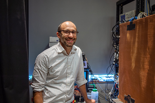
Patrick Drew led a team that found that just the pupil diameter of a mouse can determine the mouse's arousal state with high accuracy. Credit: Kelby Hochreither/Penn State
The eye is the window to the brain activity and arousal state
Researchers develop noninvasive method for determining if mice are awake or asleep
February 14, 2023
By Sarah Small
UNIVERSITY PARK, Pa. — Mice are commonly used in research to understand the brain and knowing whether the mice are asleep or awake during the experiments is critical to interpreting the results. However, because mice can transition from fully awake to fully asleep in 10-20 seconds and can sleep with their eyes open, it’s not always easy to determine their arousal state. Researchers from Penn State recently have found that just the pupil diameter can determine the arousal state of a mouse with high accuracy.
The team published their results in the Journal of Neuroscience.
“In many neuroscience lab experiments, mice perform cognitive and/or motor tasks, but they don’t always perform the tasks correctly, and it is important to understand why,” said corresponding author Patrick Drew, professor of engineering science and mechanics, of biomedical engineering, of neurosurgery and of biology.
Sleep could interfere with mice behaviors, as well as cause significant but non-task-related changes in neural activity and blood flow, according to Drew. The issue, he said, is that the sleep events can be very brief and difficult to detect.
“Previous work with sleep monitoring required many brain and muscle electrodes and extensive analysis,” Drew said. “We showed we could detect sleep events using only a video of the eye, which is technically very simple to do.”
According to Drew, the pupil is a good indicator of the arousal state of a mouse or human because its diameter is correlated with activity in a brain region called the locus coeruleus (LC). LC activity levels are important for determining alertness, including if the mouse or person wakes up from sleep.
“We found that sometimes—during rest and the kind of sleep called non-rapid eye movement (NREM) sleep—the pupil diameter was strongly anti-correlated with the amount of blood in the brain, suggesting that LC activity has a vasoconstrictory effect—meaning the blood vessels narrow in the brain when the pupil dilates,” Drew said.
To put it simply, the more dilated the pupil, the less likely the mouse is asleep.
According to Drew, these findings suggest that arousal and modulators are the main drivers of hemodynamic signals in the brain, which are used in fMRI to infer the activity of neurons. Their findings also show the sleep state of mice can be determined accurately using only a camera to monitor the eye. Monitoring the eye and pupil diameter is easier to analyze and less invasive than the standard sleep scoring that involves electrodes and other measures, and it allows easy detection of microsleep episodes during behavioral studies.
Kevin L. Turner, a recent doctoral graduate of the Penn State Department of Biomedical Engineering and the Penn State Center for Neural Engineering, and Kyle W. Gheres, a graduate of the Molecular, Cellular and Integrative Biosciences Graduate Program who was also affiliated with the Penn State Center for Neural Engineering and the Penn State Department of Engineering Science and Mechanics, are co-authors of the paper. Drew is also affiliated with the Penn State Huck Institutes of Life Sciences. The National Institutes of Health supported this work.


