


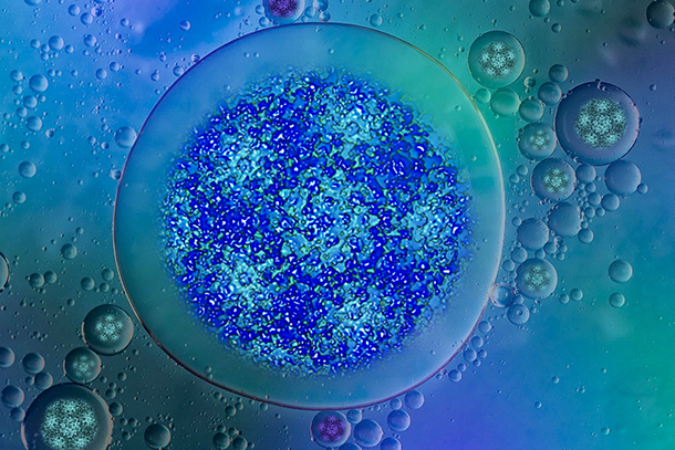
Virus particles in liquid droplets can be seen in minute detail with liquid-electron microscopes. This image, designed by the Kelly Lab, was selected as the Microscopy and Microanalysis April 2022 journal cover. CREDIT: The Kelly Lab/Penn State
New workflow, tools allow viruses to be studied in near-native environments
2/21/2022
By Mariah Chuprinski
UNIVERSITY PARK, Pa. — A study led by Penn State researchers shows new ways to view viruses and vaccine components in minute detail using liquid-electron microscopy, a new tool that allows for high-resolution imaging in near-native environments. The researchers, led by Deborah Kelly, the Penn State Lloyd & Dottie Foehr Huck Chair in Molecular Biophysics, professor of biomedical engineering and director of the Center for Structural Oncology, published their results in Microscopy and Microanalysis.
Kelly’s process allows for clearer, more detailed imaging of biological structures, such as viruses and proteins. Her findings advance current liquid-electron microscopy methods by automating processes in specimen preparation, data collection and computing, which can expedite biological research on such subjects as COVID-19 vaccine development and therapeutics.
“For the first time, we can see atomic-level features in these items in a liquid environment, as they would be contained in the human body,” Kelly said. “Typically, such items need to be frozen and are viewed in a static environment.”
The paper was selected as the cover article for the journal’s April 2022 issue. Kelly’s collaborators were William Dearnaley, technical director of the CSO; Jennifer Gray, Penn State assistant research professor in materials science; Michael Casasanta, post-doctoral fellow in biomedical engineering; biomedical engineering doctoral students G. M. Jonaid, Samantha Berry and Liam Kaylor; and industry members Madeline J. Dressel-Dukes and Michael S. Spilman.