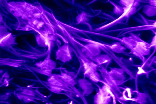
Scanning electron microscopy image showing carbon nanotubes (purple) effectively trapping Influenza viruses (light purple round objects). These trapped viruses are then analyzed by Raman spectroscopy and machine learning and they can be identified with accuracies >95%. Credit: Elizabeth Floresgomez and Yin-Ting Yeh. All Rights Reserved.
Real-time, accurate virus detection method could help fight next pandemic
June 2, 2022
By Jamie Oberdick
Editor’s note: This article originally appeared on Penn State News. Shengxi Huang, assistant professor of electrical engineering and biomedical engineering, was featured.
UNIVERSITY PARK, Pa. — A method of highly accurate and sensitive virus identification using Raman spectroscopy, a portable virus capture device and machine learning could enable real-time virus detection and identification to help battle future pandemics, according to a team of researchers led by Penn State.
“This virus detection method is label-free and not aimed at any specific virus, thus enabling us to identify potential new strains of viruses,” said Shengxi Huang, assistant professor of electrical engineering and biomedical engineering and co-author of the study that appeared today (June 2) in the Proceedings of the National Academy of Sciences. “It is also rapid, so suitable for fast screening in crowded public spaces. In addition, the rich Raman features together with machine learning analysis enable a deeper understanding of the virus structures.”
Raman spectroscopy detects unique vibrations in molecules by picking up shifts when a laser light beam induces these vibrations. To capture the viruses, a tool known as a microfluidic device would be used to trap viruses between forests of aligned carbon nanotubes.
Microfluidic devices use very small amounts of body fluids on a microchip to do medical and laboratory tests. Such a device could use virus cultures, saliva, nasal washes or even exhaled breath, including samples gathered on-site during an outbreak. The carbon nanotubes forests would filter out any foreign substance or background molecules from the host or surrounding air that could make it more difficult to get an accurate reading.
“The fact that we're using carbon nanotubes to enrich samples has been very useful because that way we are enriching the sample of viruses and eliminating other bionoise that you don't want to have when looking for a virus,” said Mauricio Terrones, Evan Pugh University Professor and The Verne M. Willaman Professor of Physics and study co-author.
Once the samples are captured and the Raman microscope examines them, then the machine learning aspect comes into play. The researchers gathered the Raman spectra of three different categories of viruses: human respiratory viruses, avian viruses and enteroviruses. This data is then used to train a machine learning model, a convolutional neural network, that identifies viruses.
"After the machine learning model is trained, then given an unknown Raman spectrum of an unknown virus, our machine learning model can automatically recognize what type of virus it is,” said Sharon Huang, associate professor of information sciences and technology and corresponding author of the study. “This includes, such as with influenza, recognizing what type it is, whether it is influenza A or influenza B, and the model can even recognize subtypes of viruses, such as H1N1 or H3N2.”
The benefits of such a device are many, according to the researchers, especially in a fast-moving outbreak.
“By providing a rapid and label-free virus detection device for virus surveillance, this approach would enable public health officials to more closely monitor the evolution of a virus,” said Yin-Ting Yeh, assistant research professor in the Eberly College of Science and co-author of the study.
Along with researchers from Penn State, George Washington University and Johns Hopkins University, researchers from the National Institutes of Health also participated in the research. The team's next steps will include collecting more Raman spectra of different human and animal viruses, including DNA viruses to grow the virus spectra database. This would enable a more extensive training of the machine learning models and enhance their ability to be generalized and to detect new strains of viruses. In addition, they will work to improve the Raman enhancement in the device to enable better signal intensities and lower bionoise levels.
“Using machine learning for Raman signal processing is not novel in itself,” said Elodie Ghedin, senior investigator for NIH’s system genomics section and co-author of the study. “What makes this approach novel is the combination of a portable virus capture device, the collection of Raman spectra from the captured viruses on this device and the rapid and accurate classification of the viruses using a machine learning model. This virus detection in real-time approach is particularly timely for tackling current and future outbreaks.”
Along with Ghedin, Terrones, Yeh, Shengxi Huang and Sharon Huang, the other Penn State authors of the study are Jiarong Ye, Ziyang Wang, Na Zhang, He Liu, Kunyan Zhang, Zhuohang Yu, Nestor Perea Lopez, Lindsey Organtini, Wallace Greene, Susan Hafenstein and Huaguang Lu. The other authors of the study are Yuan Xue, from John Hopkins, RyeAnne Ricker, from George Washington University, and Allison Roder, from NIH.
The National Science Foundation, NIH, The Huck Institutes of the Life Sciences and the Materials Research Institute at Penn State supported this research.


