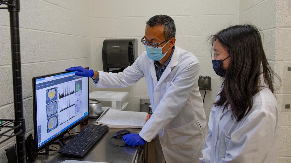
Nanyin Zhang, left, the Dorothy Foehr Huck and J. Lloyd Huck Chair of Brain Imaging in Penn State’s Department of Biomedical Engineering, looks at functional MRI and electrophysiology data with doctoral student Wenyu Tu. Using those tests, researchers will determine how rodents' brain function changes in different levels of anesthesia-induced consciousness. IMAGE: KELBY HOCHREITHER
$1.3M grant to study brain function during anesthesia-induced unconsciousness
9/30/2021
By Mariah Chuprinski
UNIVERSITY PARK, Pa. — Anesthesia has been used for surgeries and procedures for almost two centuries, but scientists are just now making advances in determining what our brains do when we lose and recover consciousness. Penn State researcher Nanyin Zhang will investigate how brain function changes during conscious and unconscious states with a four-year, $1.3 million grant from the National Institute of General Medical Sciences of the National Institutes of Health.
Zhang, the Dorothy Foehr Huck and J. Lloyd Huck Chair of Brain Imaging in Penn State’s Department of Biomedical Engineering, will use functional MRI and electrophysiology to test the brain activity of rodents while they are in different levels of consciousness. According to Zhang, the results could allow anesthesiologists to give more precise doses of the drugs in clinical settings and predict the probability of recovery for people in a coma or other vegetative states.
“We will use different doses of anesthesia to modulate the different unconscious and waking states, while we perform imaging and electrophysiological tests to trace continuous brain activity,” Zhang said.

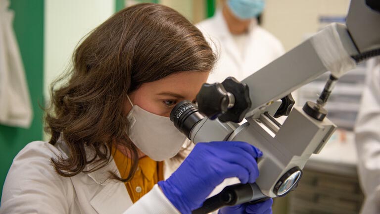
Nikki Beloate, assistant research professor of biomedical engineering, uses a microscope to determine the location of an electrode to prepare for neuronal electrophysiology testing. IMAGE: KELBY HOCHREITHER
The researchers will use the anesthetics propofol, ketamine, dexmedetomidine and isoflurane to modulate the waking and unconscious states of the rodents. Each of the drugs may have different effects on the test subjects, which Zhang said could become valuable data for clinical applications.
Zhang and his research team will pay particular attention to brain activity at the short transition point between being awake and falling deeply unconscious, as well as the point of recovering from unconsciousness, to determine brain network activity during those periods. They will compare that to the brain network organization during steady state, when researchers maintain a certain dose of anesthesia for a period of time.
Zhang and his research group, the Translational Neuroimaging and Systems Neuroscience Lab, study complex brain imaging technologies, like fMRI and electrophysiology, as well as other neuroscience technologies like chemogenetic and optogenetic methods.

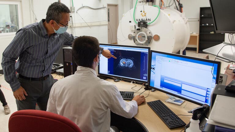
Zhang, left, and doctoral student Xu Han examine brain imaging. Behind them is the functional MRI scanner where they will perform brain function testing over the next four years. IMAGE: KELBY HOCHREITHER
Neuronal electrophysiology records the electrical activities of the brain through electrodes, which are placed either on top of the head or through a tiny needle in the brain, according to Zhang. The electrodes produce precise, detailed data akin to a “street view” of a particular region. Functional MRI, on the other hand, collects images of the entire brain, like a zoomed-out aerial photo.
“We have been trying to grasp unconsciousness for decades, and we still don’t understand it well,” Zhang said. “The brain is a complex network of neurons which do not work independently — they are tightly inter-connected.”
Zhang and his team have published five papers on this topic, but this is the first study to incorporate both electrophysiology and fMRI together as a multimodal dataset.
“The goal is to identify an imaging mechanism that can be generalized to all unconscious states, so that doctors can better understand different states of consciousness and brain activity,” Zhang said. “After almost 10 years of working on this research, it is exciting that we now have the opportunity to systematically study these concepts over the next four years.”


