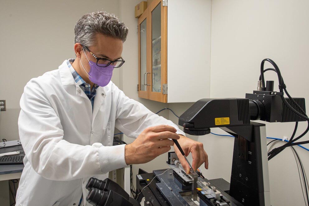
Spencer Szczesny, Penn State assistant professor of biomedical engineering and orthopaedics and rehabilitation, has a goal to develop a new way to genetically engineer synthetic tendons and ligaments. Here, Szczesny sets up a testing device mounted on a confocal microscope that will be used to evaluate the mechanical characteristics of the embryonic tendons. IMAGE: KELBY HOCHREITHER
$1.25M grant lays groundwork for synthetic tendon and ligament implants
9/23/2021
By Mariah Chuprinski
UNIVERSITY PARK, Pa. — Torn ligaments and tendons are the bane of athletes and runners, difficult to heal and often taking months or years of rehab. Currently, the only fix for severe tears is to remove intact ligaments and tendons from another part of a patient’s body, or from a cadaver, and use them to repair a knee or ankle.
This method can be costly and inefficient, according to Spencer Szczesny, Penn State assistant professor of biomedical engineering and orthopaedics and rehabilitation, but the medical field has yet to come up with a better solution. Szczesny will co-lead a four-year, $1.25 million, internationally collaborative study, with researchers from Penn State, Trinity College Dublin, Queen’s University Belfast and Dublin City University, to observe normal embryonic tendon and ligament development in chickens and mice and develop a process to replicate them synthetically.
“There is a lot of interest in developing artificial ligament and tendon replacements that integrate biologically into the bone and muscle around them and continue to live on in the body,” Szczesny said. “Our goal is to lay the groundwork by observing chick and mouse development and use that information to genetically engineer a working tendon or ligament implant.”
Earlier soft-tissue development methods, such as scaffold-based tissue engineering, have not worked clinically, Szczesny said. Early attempts in the 1970s and 1980s used synthetic polymer grafts as replacements for torn anterior cruciate ligaments, commonly known as the ACL. However, because the materials lacked the ability to remodel or heal from damage, over time the grafts stretched out, loosened up or produced plastic debris in the body and were taken off the market. Current tissue engineering techniques still utilize polymer scaffolds but also incorporate living cells, which are intended to help form an adaptive biological material around the shape of the manmade scaffold-like structure of plastic fibers.
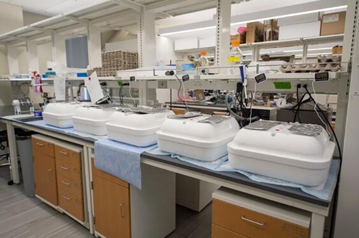
A look at the incubators that house the researchers’ fertilized eggs. The incubators control the temperature so that the chick embryos properly develop during the research. IMAGE: KELBY HOCHREITHER
“The scaffold process is fundamentally flawed because it is not how tendons form naturally,” Szczesny said. “It is actually the reverse of the way tendons develop in an embryo — cells first assemble, then build a durable structure around themselves.”
Early on in embryonic development, tendon progenitor cells line up in rows and “hold hands” in front and behind them, according to Szczesny. Once they are self-assembled, the cells start producing collagen and other fiber molecules in the gaps between them, creating long collagen fibers that connect to muscle or bone. The cells continue to produce the fibers until they become embedded in them, which together forms the larger load-bearing tendons and ligaments.
To understand this embryonic development of tendons and ligaments, Szczesny and his students, as well as co-principal investigator Paula Murphy, professor of zoology at Trinity College Dublin and an expert in developmental biology, will measure the tendons of chicks and mice at different points of development to map out the mechanical, structural and biological features at each stage.
“Chicks start to walk as soon as they hatch, which means their tendons and ligaments are already working,” Szczesny said. “So, we can really look at different developmental time points to see what is changing while the chicks develop.”
The team will measure the strength and stiffness of the developing tissue, the length, diameter and stretch of the collagen fibers, and gene expression changes, which indicate the level of biological proteins inside the cell and molecular signaling pathways. The researchers also will investigate the idea that movement inside the egg — similar to a human baby kicking in the womb — is critical to tendon development.
“Research shows that tendon cells are sensitive to movement and stimulation,” Szczesny said. “We will determine if the muscle stimulation impacts cell behavior and how they form the tissue around them. Our hypothesis is that muscle activity early on stimulates tendon and ligament cells to grip onto collagen instead of neighboring cells, forming a connection to collagen fibers, which is critical to forming a healthy tendon.”
If that hypothesis proves true, Szczesny will apply mechanical stimulation to cells when creating a tendon implant in the second phase of the project, mimicking what is happening in an egg or a human womb. The researchers also will use nanoparticles to influence tendon growth, as they can “tell” cells to turn genes on or off, according to Szczesny.
Co-principal investigator Helen McCarthy, professor at Queen’s University Belfast in Northern Ireland and expert in nanomaterial drug delivery, will lead the nanoparticle design, which should mimic the embryonic process by pushing the animals’ stem cells to release neighboring cells and bind instead to collagen fibers. If they are successful, the researchers will test the functionality of their new tendon construct in an animal model.
“This project is truly interdisciplinary, integrating international expertise in biomechanics, mechanobiology, developmental biology and materials science,” Murphy said. “We are particularly enthusiastic to bring a developmental perspective to understanding tendon biomechanics and to addressing the critical barriers that have — to date — prevented the development of functional load-bearing tendon and ligament replacements.”
Niamh Buckley, senior lecturer in personalized medicine and pharmacogenomics at Queen’s University Belfast; Aman Dhawan, Penn State associate professor of orthopaedics and rehabilitation; and Nicholas Dunne, professor of biomaterials engineering at Dublin City University, will contribute to the research.
The National Science Foundation, the Science Foundation Ireland and the Department for the Economy funded this research. The Penn State Poultry Education and Research Center is providing the fertilized eggs.


